


- 中共bat365官方网站(中国)有限公司委员会 关于预备党员转正的公示 2023-06-20
- 毕业生党员教育|公司开展“学海扬帆 见书如面”书籍漂... 2023-06-19
- 公司党委召开2023年春季学期支委学习培训会 2023-06-19
- 中共bat365官方网站(中国)有限公司委员会关于预备党员转正的公... 2023-06-19
- 中共bat365官方网站(中国)有限公司委员会关于预备党员转正的公... 2023-06-19
- 中共bat365官方网站(中国)有限公司委员会关于预备党员转正的公... 2023-06-19
- 中共bat365官方网站(中国)有限公司委员会关于确定程博等同志为... 2023-06-14
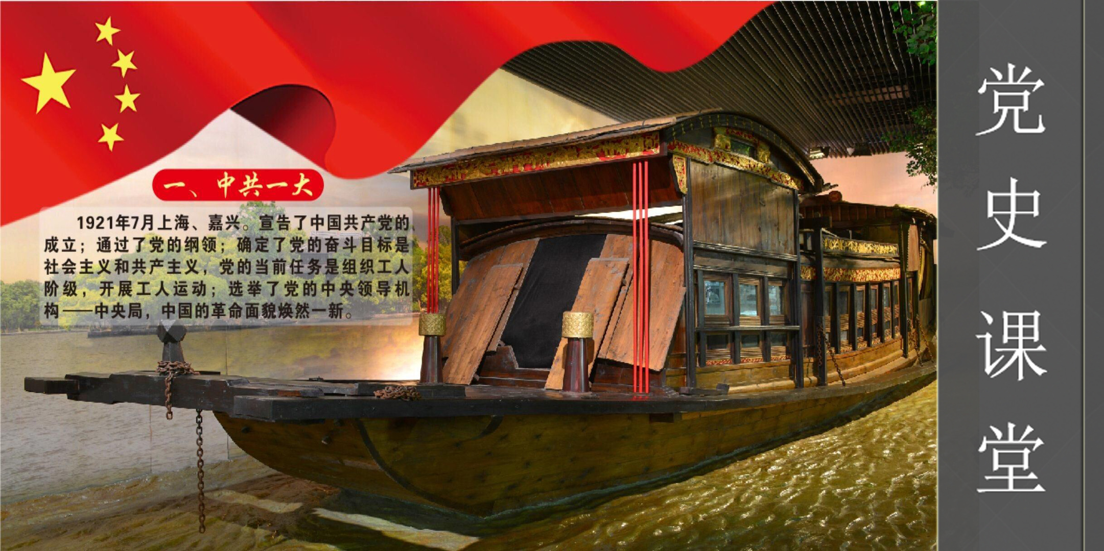
党建工作
>
教学管理
>
- bat365官方网站(中国)有限公司 2023年度bat365官方网站“老员工创新... 2023-05-31
- bat365官方网站2023年春季学期全日制研究生学位答辩安排... 2023-05-10
- bat365官方网站(中国)有限公司 2023年bat365官方网站研究生科研创新... 2023-05-19
- bat365官方网站(中国)有限公司 2023年重庆市研究生科研创新项... 2023-05-16
- bat365官方网站公共管理、行政管理2023年春季学期学位论... 2023-05-11
- bat365官方网站 重庆市2023年“课程思政”示范项目推荐... 2023-05-08
- bat365官方网站(中国)有限公司2023年哲学博雅班入选名单公示 2023-05-08
员工工作
>
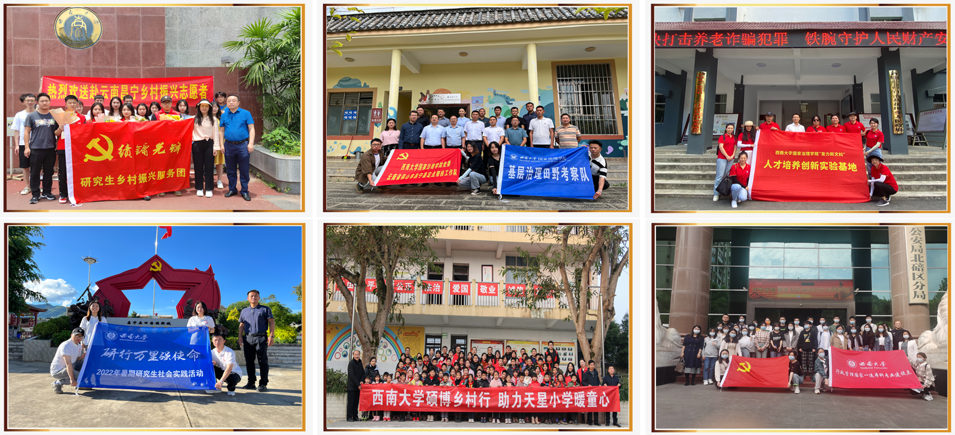
- 2023年暑期“三下乡”|公司实践团与万盛万东镇联合开... 2023-07-10
- 2023年暑期“三下乡”|社会实践团举办“传统文化进社... 2023-07-10
- 2023年暑期“三下乡”|社会实践团开展生命教育系列活... 2023-07-10
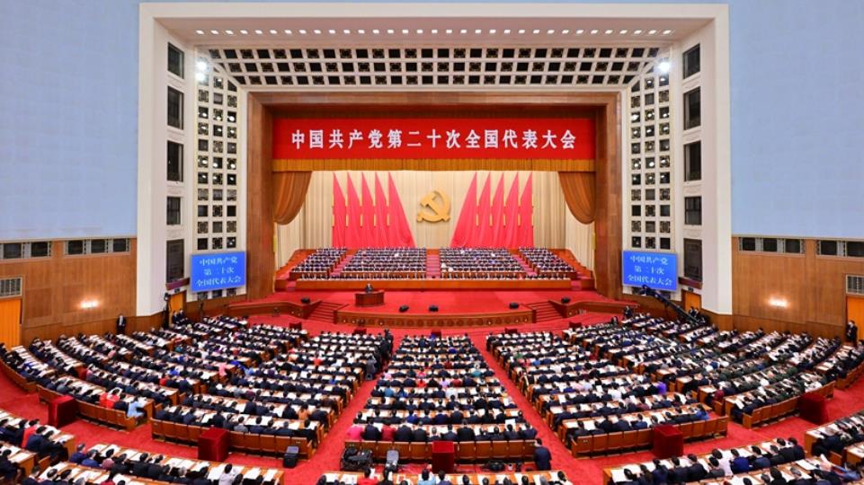
- 2023年暑期“三下乡”|社会实践团举办“学习二十大,... 2023-07-07
- 2023年暑期“三下乡”|“初生朝阳,雏鹰起飞”儿童素... 2023-07-07
- 2023年暑期三下乡|“关爱生命,友好童行”成长训练营... 2023-07-07
- bat365官方网站“我身边的诚信故事” 主题征文作品评选... 2023-06-29
人才招聘
>
- bat365官方网站2024年硕士研究生入学考试参阅书目 2023-06-30
- bat365官方网站2023年“中国-希腊文明比较”专项博士学位研... 2023-05-30
- bat365官方网站(中国)有限公司关于2023年博士研究生招生拟录取... 2023-05-22
- bat365官方网站2023年“中国-希腊文明比较”专项博士学位研... 2023-05-19
- bat365官方网站2023年“中国-希腊文明比较”专项博士学位研... 2023-05-19
- bat365官方网站2023年“中国-希腊文明比较”专项博士学位研... 2023-05-12
- bat365官方网站关于2023年公共管理(MPA)硕士研究生拟... 2023-04-27
社会服务
>
- 重庆市社区戒毒社区康复工作人员2020年综合能力提升专... 2020-12-15
- 重庆市璧山区2020年乡村振兴和扶贫开发专题培训班在我... 2020-12-14
- 重庆市发改委2020年新提任处级领导干部和新进人员综合... 2020-11-24
- 重庆市发展和改革委员会2020年新提任处级领导干部和新... 2020-11-18
- 重庆市林业局2020年处级领导干部综合能力提升培训班在... 2020-11-10
新媒体矩阵
-
bat365官方网站(中国)有限公司
-

逻辑与智能研究中心
-
西大群学
-
重庆文化产业bat365官方网站研究院